Novel instrument could transform research at UCI
Mass cytometer expands cell profiling capabilities of scientists across campus
Imagine looking up at the night sky and trying to find the Big Dipper but only being able to see one star at a time. You wouldn’t recognize the constellation’s familiar pattern. This is analogous to the problem solved by mass cytometry technology. By allowing researchers to observe and analyze an increased number of data points on a single-cell basis, mass cytometry aids in the resolution of the biological picture they’re studying.
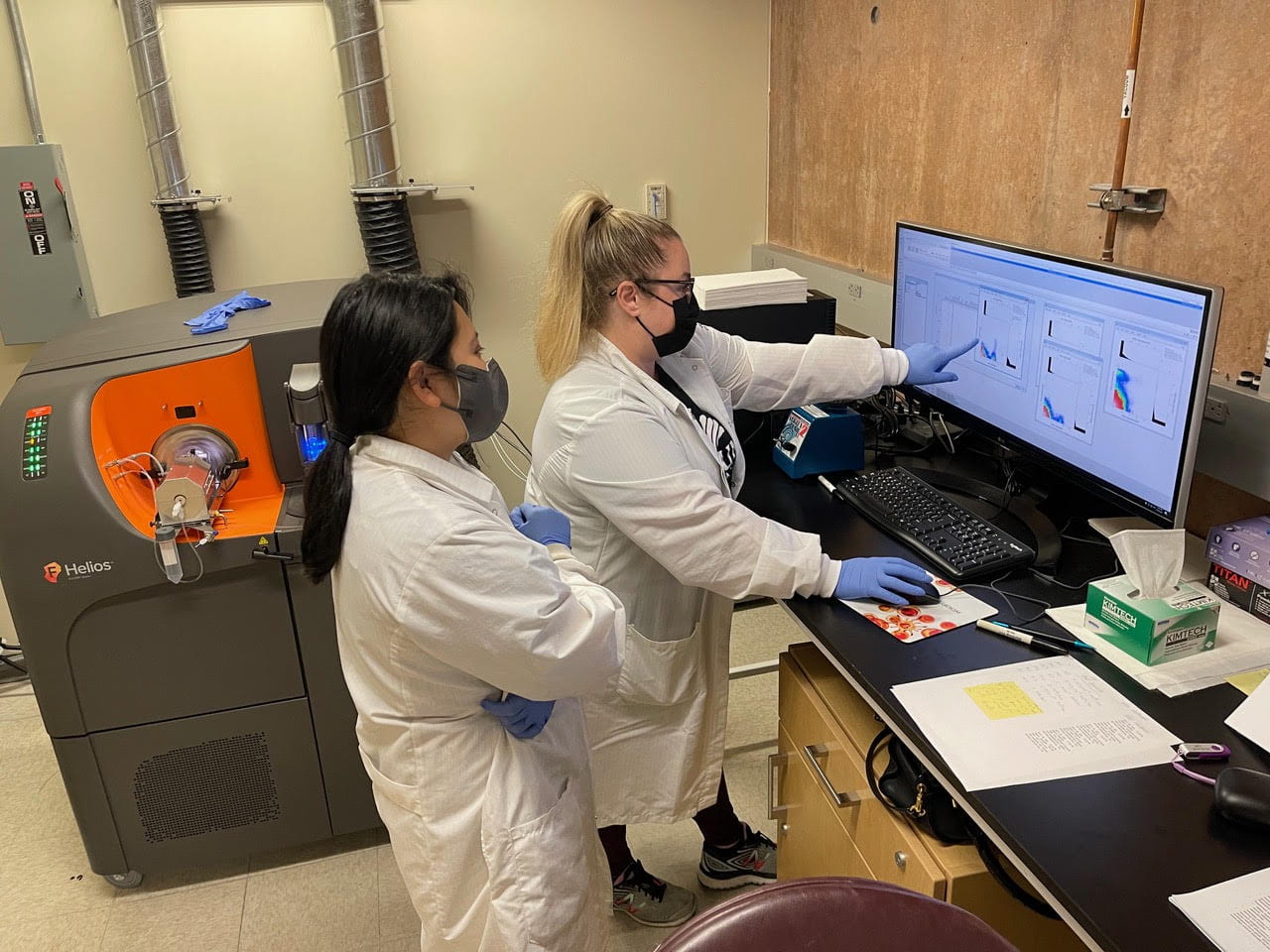
In partnership with the schools of medicine and biological sciences, the Office of Research and the UCI Health Chao Family Comprehensive Cancer Center – and with a generous gift from the William and Ann Cullen Endowment – UCI’s Sue & Bill Gross Stem Cell Research Center is home to a new mass cytometer. A powerful instrument that can analyze a high number of proteins in an individual cell, it is the only one of its kind in Orange County and just the third on a University of California campus. The Helios mass cytometer’s multiparameter analysis capability has the potential to transform research by providing insights not previously possible.
Understanding how proteins in cells function and interact is key to discovering treatments for conditions ranging from Alzheimer’s disease and COVID-19 to multiple sclerosis and traumatic brain injuries. The greater the number of proteins that can be simultaneously measured, the deeper the understanding scientists can gain into the locations, relationships and communication mechanisms of different cell types.
“Until recently, these measurements were limited in number, ranging from 10 to 20 depending on the instrument used. The Helios mass cytometer allows researchers to profile more than 50 at one time, and this data has the potential to reveal changes that can’t be measured with other technology,” says Vanessa Scarfone, flow & mass cytometry core manager at the Sue & Bill Gross Stem Cell Research Center. “More detailed information aids in understanding complex cell functions and associations, and that’s critical for developing targeted treatments for many diseases.”
The Helios combines flow cytometry, a technology that rapidly measures and analyzes cell characteristics as they flow past single or multiple lasers while suspended in a buffered salt-based solution, with mass spectrometry, an analytic technique that identifies cells’ protein substances. The new tool generates proteomic data in higher resolution and dimensionality than traditional fluorescence-based systems.
Research applications
The Helios is currently being used by faculty members from across campus for research on cancer, wound healing, inflammatory myopathies, neuroinflammation and stem cells.
Chris Halbrook, Ph.D., assistant professor of molecular biology & biochemistry, is working to reprogram the microenvironment that surrounds and feeds pancreatic cancer tumors to reduce immune suppression and enable immunotherapy. His team conducted the first experiment on the Helios. “We wanted to track how primary and metastatic pancreatic tumors change in response to therapeutic interventions,” Halbrook says. “After analyzing our results, we identified unique differences in the immune response in tumors obtained from the pancreas and liver. I think this data demonstrates the power of high-dimensional flow technology.”
Maksim Plikus, Ph.D., a professor of developmental & cell biology who’s exploring wound healing, says, “The skin is a complex network of many cell types. The Helios will allow me to examine specific proteins from these cells and how they aid in the wound healing process.”
Thomas Lane, Ph.D., Chancellor’s Professor of neurobiology & behavior, and Craig Walsh, Ph.D., professor of molecular biology & biochemistry, are studying demyelination in multiple sclerosis. The loss of myelin, a type of insulating tissue that surrounds and protects nerves throughout the body, causes neurological deficits such as vision changes, impaired coordination, fatigue and cognition problems.
T cells are a particular type of white blood cells that are part of the body’s immune system and develop from stem cells in the bone marrow. Matthew Inlay, Ph.D., associate professor of molecular biology & biochemistry, is investigating how T cells respond to drug treatments that could help prevent graft-versus-host disease. This condition occurs in bone marrow transplant patients when T cells in donated stem cells attack healthy cells.
“The Helios enables deep profiling across a range of cells and can be applied to many areas of research,” Scarfone says. “Our facilities are available to UCI faculty, as well as scientists around Southern California.”
Publicly launched on Oct. 4, 2019, the Brilliant Future campaign aims to raise awareness and support for UCI. By engaging 75,000 alumni and garnering $2 billion in philanthropic investment, UCI seeks to reach new heights of excellence in student success, health and wellness, research and more. The Sue & Bill Gross Stem Cell Research Center plays a vital role in the success of the campaign. Learn more by visiting https://brilliantfuture.uci.edu/uci-school-of-medicine.